

 Click it and Unblock the Notifications

Latest Updates
-
 Purported Video of Muslim Mob Lynching & Hanging Hindu Youth In Bangladesh Shocks Internet
 -
 A Hotel on Wheels: Bihar Rolls Out Its First Luxury Caravan Buses
 -
 Bharti Singh-Haarsh Limbachiyaa Welcome Second Child, Gender: Couple Welcome Their Second Baby, Duo Overjoyed - Report | Bharti Singh Gives Birth To Second Baby Boy | Gender Of Bharti Singh Haarsh Limbachiyaa Second Baby
 -
 Bharti Singh Welcomes Second Son: Joyous News for the Comedian and Her Family
 -
 Gold & Silver Rates Today in India: 22K, 24K, 18K & MCX Prices Fall After Continuous Rally; Check Latest Gold Rates in Chennai, Mumbai, Bangalore, Hyderabad, Ahmedabad & Other Cities on 19 December
 -
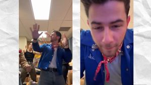 Nick Jonas Dancing to Dhurandhar’s “Shararat” Song Goes Viral
 -
 From Consciousness To Cosmos: Understanding Reality Through The Vedic Lens
 -
 The Sunscreen Confusion: Expert Explains How to Choose What Actually Works in Indian Weather
 -
 On Goa Liberation Day 2025, A Look At How Freedom Shaped Goa Into A Celebrity-Favourite Retreat
 -
 Daily Horoscope, Dec 19, 2025: Libra to Pisces; Astrological Prediction for all Zodiac Signs


A Neurosurgeon Explains About Orbital Tumors: Diagnosis, Treatment & Management
Tumors and inflammations can occur behind and around the eye. An orbital tumor refers to a tumor positioned in the 'orbit', which is the bony socket in the front of the skull that comprises the eye. Even a miniscule tumor in this small, crowded region can cause significant symptoms and functional effects. Eyes bulge forward and cause serious vision problems when the tumors are large in size. Orbital tumors are both benign and malignant in nature.

Among children this tumor may be apparent at birth or acquired later. Although most orbital tumors are benign, malignancies such as retinoblastoma and rhabdomyosarcoma are vision- and life-threatening. In adults, the majority of orbital tumors include cysts, vascular lesions (arising from blood vessels), lymphomas, neurogenic tumors (arising from nerves), and secondary tumors (either metastatic or spread directly from the surrounding sinuses or cranium).
Eyes and the structures around it (orbit) is the second most complex organ only next to the brain. Any abnormal growth of this tissue is either solid or cystic present with a spectrum of signs and symptoms depending on age of the patient and biological nature of the tumor.
Signs & Symptoms
- Bulging of the eye
- Double vision
- Loss of vision
- Droopy or swollen eyelid
- Feeling of pressure in the eye
- Flattened eyeball
- Pain and inflammation
On occasions these tumors are incidentally detected while imaging the head for other conditions and patients may develop their symptoms over time. Rapidity of onset and the location and nature of their symptoms are often important clues to determine whether a problem is benign or cancerous. . As the eyes are pushed forward, the eyelids often appear to be retracted from it hence patients develop bulging of the eye (proptosis or exophthalmos) and as larger tumors displace the axis of the eye, patients may see double vision. Some tumors can actually be seen or felt on examination. Those tumors arising from the optic nerve present with progressive diminution of vision. It is therefore important to identify ocular and orbital tumors early.
Diagnosis
Determining orbital tumors can be done through using advanced diagnostic procedures and technology to efficiently diagnose, carefully observe the condition. Diagnostic procedures include:
1. Biopsy: A sample of the tissue is removed by a small incision by an expert physician, this tissue is used examine the type of tumor present.
2. Computerized tomography (CT) scans: X-rays and computers are used in the test to create images of the tumor to detect its location, size and various other abnormalities.
3. Magnetic resonance imaging (MRI): This particular test uses a powerful magnet and radio waves to create images of the tumor to see its size and location, and to rule out other deformities.
Though Imaging studies have rapidly evolved in last few decades ultrasonographic examination of the orbit is still helpful in the evaluation of cystic lesions and angiography is still the imaging modality of choice for vascular lesions, such as ateriovenous malformations and low-flow ateriovenous fistulas and Cerebral angiography should be undertaken in any patient with pulsatile exophthalmos. Magnetic resonance imaging gives high-resolution images of the standard components of the orbit and of nonosseous lesions in three dimensions. In cases of osseous lesions, CT is the modality of choice, either alone or in combination with an MR imaging.
In early stages significant percentage of these tumors are treated by the ophthalmologist alone, but in view of it complex anatomical entity, management of orbital tumors poses a challenging surgical task, needing proficiency with a multitude of approaches, hence it is desirable to deal these tumors by multidisciplinary team approach, undertaken by well-experienced neurosurgical team along with oculoplastic surgeon for tumors involving the orbital cranial junction and the superior orbital compartment to facilitate optimal removal of the tumor and skull base sealing as well as good cosmetic results. And involvement of a radiation oncologist to the team for adjuvant radiotherapy/chemotherapy would be beneficial for prolonged recurrence free and better quality of life. However, not all tumors require surgical excision and in some, radiation, chemotherapy, or immunotherapy may be the indicated form of treatment.
The article has been contributed by Dr. Amaresh S Bhaganagare, Consultant Neurosurgeon at HCG Cancer Hospital Bengaluru
Disclaimer: The information provided in this article is for general informational and educational purposes only and is not intended as a substitute for professional medical advice, diagnosis, or treatment. Always seek the advice of your physician or a qualified healthcare provider with any questions you may have regarding a medical condition.


