

 Click it and Unblock the Notifications

Latest Updates
-
 Purported Video of Muslim Mob Lynching & Hanging Hindu Youth In Bangladesh Shocks Internet
 -
 A Hotel on Wheels: Bihar Rolls Out Its First Luxury Caravan Buses
 -
 Bharti Singh-Haarsh Limbachiyaa Welcome Second Child, Gender: Couple Welcome Their Second Baby, Duo Overjoyed - Report | Bharti Singh Gives Birth To Second Baby Boy | Gender Of Bharti Singh Haarsh Limbachiyaa Second Baby
 -
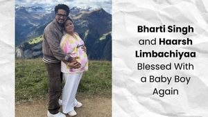 Bharti Singh Welcomes Second Son: Joyous News for the Comedian and Her Family
 -
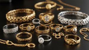 Gold & Silver Rates Today in India: 22K, 24K, 18K & MCX Prices Fall After Continuous Rally; Check Latest Gold Rates in Chennai, Mumbai, Bangalore, Hyderabad, Ahmedabad & Other Cities on 19 December
 -
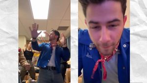 Nick Jonas Dancing to Dhurandhar’s “Shararat” Song Goes Viral
 -
 From Consciousness To Cosmos: Understanding Reality Through The Vedic Lens
 -
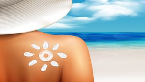 The Sunscreen Confusion: Expert Explains How to Choose What Actually Works in Indian Weather
 -
 On Goa Liberation Day 2025, A Look At How Freedom Shaped Goa Into A Celebrity-Favourite Retreat
 -
 Daily Horoscope, Dec 19, 2025: Libra to Pisces; Astrological Prediction for all Zodiac Signs


Bone Mineral Content and Osteoporosis
To detect the presence of early osteoporosis, the mineral content of bone is measured. It helps predict sites at risk for fracture, and monitor the course of disease and response to therapy. Bone mineral content is the amount of bone mineral divided by the bone-scanned area. The size and density of skeletal bone and bone mineral content are interdependent. Difference in bone mineral content may reflect a different in either bone size or bone density. Small skeleton may results in a low bone mineral content.
The index BMC/bone area is commonly called bone mineral density (BMD),
What is osteoporosis?
Osteoporosis is a systemic skeletal disease characterized by low bone mass and micro architectural deterioration of bone tissue, with a consequent increase in bone fragility and susceptibility to fracture risk. Those who achieve a higher peak bone mass are less at risk of suffering from osteoporotic fracture, later in life. Quantitative assessement of bone mineral content helps diagnosis the disease. Bone mineral density is the determinants of bone strength. By measuring bone mineral density, it is possible to predict the risk of fracture.
The amount of calcium in regions of bones is measured by bone mineral density. Methods for measuring bone mineral content are fast, non-invasive, painless and available on an outpatient basis.
Taking dual energy x-rays (DEXA) or CT scan (Osteo CT or QCT) of bones in the spinal column, wrist, arm or leg are methods of measuring bone mineral density. By comparing the numerical density of the bone with empirical (historical) data bases of bone density, it is possible to diagnose osteoporosis
Measuring
Bone
Mineral
Density
Dual
Energy
X-ray
Absorptiometry
(DEXA)
is
a
widely
accepted
method
of
measuring
bone
mineral
density.
This
method
requires
no
injections,
invasive
procedures,
sedation,
special
diet
or
any
other
advance
preparation.
It
will
take
only
few
minutes
to
complete
the
exam.
Bone mineral content can be measured using ultrasound. This is a newly developed method. This method measures bone mineral content at the patient's heel. It is less expensive.
Different types of Bone Mineral Density Tests
- Ultrasound measures the heel
- DEXA (Dual Energy X-ray Absorptiometry) measures the spine, hip or total body
- SXA (single Energy X-ray Absorptiometry) measures the wrist or heel
- PDXA (Peripheral Dual Energy X-ray Absorptiometry) measures the wrist, heel or finger
- RA (Radiographic Absorptiometry) uses an X-ray of the hand and a small metal wedge to calculate bone density
- DPA (Dual Photon Absorptiometry) measures the spine, hip or total body
- SPA (Single Photon Absorptiometry) measures the wrist
- QCT (Quantitative Computed Tomography) measures spine or hip
Who needs Bone Mineral Density Measurements?
- Post-menopausal women with at least one additional risk factor (other than menopause).
- All women older than 65 regardless of risk factors.
- Post-menopausal women who present with fractures.
- Women considering therapy for osteoporosis, if bone mineral density (BMD) testing would affect the decision.
- Women who have been on hormone replacement therapy (HRT) for prolonged periods of time.
Disclaimer: The information provided in this article is for general informational and educational purposes only and is not intended as a substitute for professional medical advice, diagnosis, or treatment. Always seek the advice of your physician or a qualified healthcare provider with any questions you may have regarding a medical condition.


